Applying Task Observations
to Improve MRI Presentation
School of Computing Science
Simon Fraser University
Burnaby, BC V5A 1S6 Canada
+1 604 268-6605
|
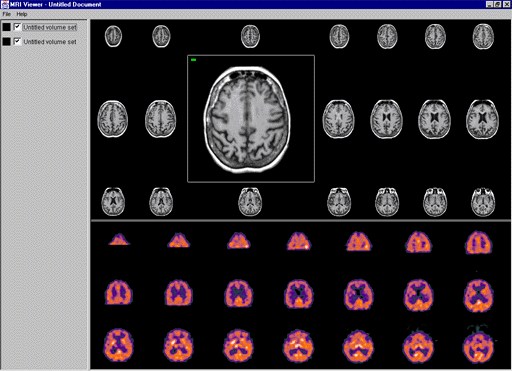
ABSTRACT In Magnetic Resonance Imaging (MRI), anatomical structures are visualized by scanning successive slices of the human body. Traditionally, radiologists use a large light screen display to view MRI volume sets. This paper describes a video mock-up and a resulting software prototype developed to display MRI volume sets on a traditional computer monitor. Five key objectives were identified concerning the design of the software prototype. Keywords Medical imaging, user interfaces, magnetic resonance imaging, fish-eye visualization, prototyping INTRODUCTION During the process of magnetic resonance imaging (MRI), a huge tube-shaped scanner creates anatomical images of a specified segment of the human body. Traditionally, a large light screen display is used to present a series of images referred to as a volume set. These volume sets may contain many images and up to eight volume sets may be on the light screen at the same time. However, the costs for buying and maintaining light screens are high and films tend to get lost. Furthermore, some radiologists have developed allergies to the material in the films. A current trend in radiology is on-line medical imaging. By maintaining images on-line, more complicated manipulations can be performed on the digital images. The data is also not restricted to one location. This is important for telemedicine where images are transferred over a network for diagnosis and educational purposes. Moving from the large light screen to a desktop computer monitor means that the available space is tremendously reduced. Many of today’s medical imaging systems only display a subset of the required images. Therefore, contextual information is lost. This is important because contextual information is crucial for diagnosing abnormalities. This paper presents an implementation of a new approach to MRI viewing on a desktop computer. We discuss previous research which highlighted the requirements for our design and five key objectives of our system. PREVIOUS WORK Van der Heyden [1] explored the "screen real-estate" problem from the perspective of MRI analysis. This research demonstrated the importance of providing both detailed and contextual information. The use of fish-eye visualization techniques was proposed to provide detail through magnification of the focus and maintain context through compression. In addition, the traditional light screen metaphor was applied to facilitate the transition to desktop systems. In particular, volume sets are individually contained in windows with rows and columns of images. Each gray-scale image is presented on a white background. A preliminary study was conducted at UBC Hospital, Vancouver, to investigate the proposed approach. The results from the study showed that images of at least 30 x 30 pixels were adequate to distinguish between images while accurate diagnosis demands full resolution (256 x 256 pixels for MRI). The radiologists also found the excessive white space between images to be very distracting. Even when using the traditional light screen, they sometimes put up extra films to minimize the bright white space or switch off the light from behind. Extending the Study The next phase of this research project involves development of software to provide radiologists with an interactive environment. This paper describes two initial activities: a video mock-up of the interface and a prototype of the software. Video Mock-Up In order to illustrate the proposed interface, video and paper-based prototyping techniques were used. We selected three scenarios that were representative of key tasks within MRI analyses. These scenarios were demonstrated using the paper-based interface and videotaped. This mock-up was used to solicit feedback and gather information to assist in the design process for the software prototype. Software Prototype There were five key objectives for the software prototype of the MRI viewer. First, to present one or more volume sets in a single window. Second, to utilize fish-eye visualization techniques to provide detail and context. Third, to alleviate the distraction caused by white space between images. Fourth, to create an environment that allows for extensibility. Fifth, to integrate data collection techniques. Figure 1 shows a screenshot of the MRI viewer.
Figure 1. Screenshot of the MRI viewer Presentation of Volume Sets The MRI viewer allows for the display of one or more volume sets. Volume sets are loaded into memory and selected for display. The program lists the currently loaded volume sets and whether or not they are currently visible. When more than one volume set is selected for display they are stacked vertically. A thin bar separates the different volume sets. The bar can be dragged up or down to provide more space for a volume set. Fish-Eye Visualization In order to provide both detailed and contextual information, we chose a fish-eye visualization technique that preserves orthogonality without distorting the images [2]. When the mouse pointer is moved over an image, a dark gray bounding box appears to indicate which image will be manipulated. Clicking the image magnifies it. Scans that are not magnified shrink to at least 30 x 30 pixels. Magnified scans try to attain their full resolution. A green box in the corner of an image indicates that it has reached full size. A red box means the image is smaller than that. Removal of White Space In response to the radiologists’ comments from the preliminary study, the background color was chosen to be black. This should help reduce the distraction previously noted using the white background. Nonetheless, functions were included to create user-defined background colors if desired. Extensibility The software was implemented using Java and object-oriented techniques to provide for easy extensibility and platform independence. New image layout techniques are easily plugged into the program by deriving from the class that manages the display. This also applies to other display objects, for example, to integrate annotated images. Data Collection All classes contain three data collection functions: Log, Error, and Record. These functions are used to trace user actions during a user study. Messages sent to these functions are pipelined to the Java applet where they can be saved in a file. FUTURE WORK As this is the first version of the MRI viewer, it is clear that the program is still in a growing and maturing phase. There are several potential extensions that will be considered.
CONCLUSION We presented a new approach for medical image viewing and implemented a software prototype. This will help get more precise feedback on our design approach from users of the software, specifically radiologists. Most radiologists cannot afford to spend a large amount of time learning to use an application. We are currently using this software in our ongoing collaborations with local radiologists. Acknowledgements I would like to thank Dr. Kori Inkpen and Dr. Stella Atkins for their help and guidance with this project. Thanks also to Garth Shoemaker and Yingchen Yang for help with the video mock-up. REFERENCES |
