My Master’s Thesis Research
The M.Sc. program at Simon Fraser University is typically a two year program and includes five graduate courses plus research. I completed all course work in half a year and then worked on the research project described here. Working on this project, I gained practical experience with techniques such as field observations, contextual inquiry, rapid prototyping, user studies, statistical and qualitative analysis.
Abstract
Keywords: Human-Computer Interaction, Graphical User Interfaces, Visualization, Medical Imaging
Due to the continuous drop in computer hardware prices, the use of high-end computer systems has become attractive to many hospitals. Radiology departments are now facing the transition from the use of traditional light screens and photographic films to online medical imaging systems. These new systems offer several advantages over traditional methods: films are less likely to be lost, automatic anomaly detection can improve diagnosis, and high-speed networks allow for cross-site consultations (telemedicine). However, the use of desktop monitors severely limits the space in which medical images can be viewed. This applies particularly to Magnetic Resonance Imaging (MRI) where, frequently, up to eight films, each containing up to 20 images, are viewed simultaneously on a light screen. As a result, the screen real-estate problem inherent in desktop monitors becomes a serious restriction for the radiologists.
In a previous requirements analysis, researchers suggested displaying selected images in full resolution with the surrounding images remaining on the screen although reduced in size (called detail-in-context). Based on feedback obtained from this previous research, and from our additional observations of radiologists in their workplace, we have extended the algorithm of this detail-in-context technique. We have implemented the extended technique in a software system that allows users to navigate sequences of images such as those found in MRI and in other types of image sequences such as in Meteorology, Video Editing, and Animation.
To evaluate our system, we conducted a user study with university students. The detail-in-context technique was compared to an implementation of the thumbnail technique which is utilized in many commercially available medical imaging systems. Results show that performance as well as user preference was similar for both display techniques. Our analysis of the computer logs recorded during the study, however, suggests that the detail-in-context technique can accomodate a variety of individual strategies and offers strong comparison capabilities. The thumbnail technique, on the other hand, strongly encourages a sequential examination of the images but allows for higher magnification factors. This research has implications on the selection of appropriate display techniques in many areas dealing with image sequences, including radiology.
Screenshots
The experiment involved two pieces of software. Both presented an image sequence and provided mechanisms to navigate it. One of them was an implementation of a detail-in-context technique. In the other implementation, thumbnails were used to select images for magnification.
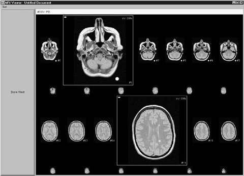
Detail-in-context techniques display information on multiple magnification levels. In this example, selected images are displayed at high magnification factors. To provide contextual information, unselected images remain around the selected ones at lower magnification factors.
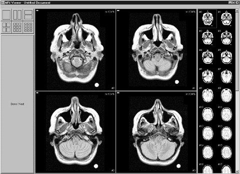
This is a screen shot of the thumbnail implementation. The thumbnail bar on the right side of the screen displays small versions of the images. Clicking on an image causes it to be magnified in the large display area to the left of the bar. Four successive images are displayed in that area by default but different layouts can be chosen by clicking on the buttons at the top left corner of the screen.